Radiation Oncology
Last Update: 07/10/2024
Radiotherapy treatment
Radiotherapy treatment is a treatment method aimed to eliminate tumor cells by irradiating the diseased area by means of radiation therapy devices. Treatments are done in daily sessions and continue over a period of several days to weeks. The radiation oncologist doctor decides on the duration and form of treatment.
Radiotherapy treatment (beam therapy – water treatment) is applied alone or in combination with surgery / chemotherapy in cancer disease. When radiotherapy is planned and implemented by a radiation oncology doctor with the support of computerized planning, the success rates increase. Therefore, the combination of technology and treatment team is important in radiotherapy treatment.
Radiotherapy treatment is a treatment method used in approximately half of cancer patients today. 3D conformal radiotherapy planning, radiotherapy using IMRT applications is effective in the treatment of cancer disease.
The contribution of technological innovations to radiotherapy:
Radiotherapy treatment is constantly updated in line with technological developments. Tumor areas can be treated without harming the surrounding organs through treatment planning systems (TPS) with applications under the supervision of a radiation oncologist. Human factor and technological advances help the treatment of patients with coordination.
As a result of the detailed examination of the diseased area with a computerized screening method, a distinction can be made between the diseased area and the normal area. In this way, radiation therapy can only be applied to the tumor area and side effects can be reduced.
Separate and Customized Treatment for Each Patient:
As a result of the reports and films examined by your doctor, a treatment plan specific to the patient emerges. This detailed treatment plan prepared by the radiotherapy team provides gains in the treatment of the disease with the daily controlled treatment practices.
Computerized planning support is used in treatment practices in our clinic. Treatment planning and implementation is carried out as a team with radiation oncologist, radiation physicist and radiotherapy technicians.
Services are provided free of charge within the scope of Private Health Insurance and SSI.
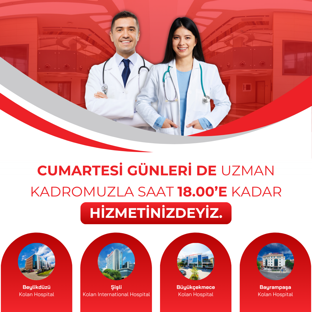
Şişli Kolan International Hospital Radiotherapy Center
Kaptanpasa Mah. Darülaceze Cad. No: 14 Okmeydanı / Şişli / İSTANBUL
0212 222 0 888
Patient consultation number and Whatsapp line 0530 955 08 64
PET / CT
PET (Positron Emission Tomography) is a functional imaging method that provides physiological information required for clinical diagnosis based on changes in tissue metabolism. The most important advantage of PET is the use of positron emitting biological radioisotopes that mimic the natural components in the body. (Such as Carbon-11, Oxygen-15, Nitrogen-13, Flor-18)
PET shows the distribution of the follower molecule (usually a sugar component F-18-FDG) marked with a specific radiopharmaceutical to the body in all organs. Radioactive followers concentrate on the areas that use it most, that is, around the tumors. During the glucose metabolism period, F-18 breaks down and emits positrons perceived by the PET unit. At the same time, the ring-shaped CT unit produces high-resolution 3D X-ray images of the part of the body being examined. The result is a combined ‘anatomo-molecular’ image showing the location, size, metabolism, and spread of tumors. Anatomical detail and functional information are obtained in a single image.
Although PET-CT is used in the most common oncology, it also enables early diagnosis in neurological diseases (dementia, epilepsy, alzheimer, etc.) and cardiology.
PET – CT, lung cancer, mesothelioma, lymphoma, melanoma, head – neck, small intestine, large intestine-colorectal, esophageal cancers, breast, thyroid and other endocrine tumors, pancreas, liver, kidney, bladder, ureter, testicle, penis and it is used in other male genital, gynecological, brain, musculoskeletal system tumors.
Usage areas
- In determining the extent of the disease (staging) before treatment,
- In determining the true tumor tissue in patients who will receive radiotherapy (radiation therapy), planning treatment, irradiation of the right area at the right dose,
- In evaluating the response to treatment (investigation of tumor response after chemotherapy or radiotherapy),
- In evaluating the effectiveness (chemosensitivity) of chemotherapy applied,
- Staging after treatment and evaluating the effectiveness of treatment,
- Re-staging of patients with recurrence,
- It is used to investigate the primary focus in cancers with metastases but no tumor focus.
Matters Needing Attention
- One day before the shooting, the patient should rest and not do things that require physical activity.
- Sugary foods and beverages (fruit, juices) should not be consumed. A minimum of 6 hours of fasting is required before the examination. The patient should not get cold while coming to the examination especially in winter.
- After the preliminary preparations on the day of the examination, the patient’s blood sugar is checked.
- FDG molecule (radioactive substance) labeled with Flor-18 which is a sugar derivative and positron irradiation is administered to the patient through the vascular access.
- It is waited between 40-60 minutes for the drug to be fully spread throughout the body. During the waiting period, the patient should rest, not move and not speak unless it is very necessary.
- After the waiting period is completed, the patient is taken to the drawing room.
- In a total of 12-20 minutes, both computed tomography and PET images are obtained in three dimensions in the same session.
RADIATION ONCOLOGY
Radiation oncology; It means the irradiation of cancerous tissue. Radiotherapy treatment aims to eliminate tumor cells by irradiating the diseased area through advanced technological treatment devices.
Treatments are done in daily sessions and can last from a few days to a few weeks.
Radiotherapy is a team job. In the Kolan Hospital Group Radiation Oncology Department, the treatment prepared and planned by an experienced team of radiation oncology specialists, radiophysics specialists, radiotherapy technicians and dosimetrists is applied. It is the responsibility of the Radiation Oncology specialist to decide what dose and how long the tumor tissue will be irradiated, and to follow the side effects that may occur in the early and long term during radiotherapy. Other members of the team are responsible for the regular administration of the planned treatment to the patient every day and for the follow-up of the patient.
Radiotherapy treatment is a successful treatment that is used successfully in a significant part of cancer patients today.
Radiotherapy treatment using advanced radiotherapy techniques, 3D radiotherapy planning, IMRT (intensity-adjusted radiotherapy) applications is effective in the treatment of cancer disease.
IMRT (Intensity Adjusted Radiotherapy) in Head, Neck and Brain Tumors
If head and neck tumors can be diagnosed at an early stage, they are usually treated by surgical method. If the tumor has spread to the lymph nodes and there are other high risk factors, radiotherapy is also used in the postoperative period. IMRT is a major factor in the success of this treatment.
Radiotherapy is applied alone in some cases, but often in combination with chemotherapy.
Diseases using IMRT
- Early stage vocal cord tumors
- Head and neck tumors
- Brain tumors
- Nasopharyngeal cancer
- Cancer of the esophagus (esophagus)
Today, thanks to modern radiotherapy techniques and devices, irradiation is considerably reduced. With three-dimensional and intensity-adjusted radiotherapy, high doses of radiation can be given to the cancerous area, while low doses can be given to the tissues and organs around the cancerous area, that is, the damage in these areas can be minimized.
For detailed information 444 1 443
Radiation Oncology Doctors